By Dr. Anup Ramani

Kidney stones can cause excruciating pain and, if left untreated, can lead to severe complications. One of the most common procedures for removing kidney stones is ureteroscopy (URS), a minimal invasive surgery that involves using a scope to remove stones from the kidney or ureter. During URS kidney stone removal, a stent is often placed in the ureter to keep it open and promote healing after the procedure. However, the placement of the stent is not a straightforward task. There are several technical considerations that need to be taken into account to ensure the safety and comfort of the patient during and after the procedure.

In this article, we will explore the technical considerations involved in stent placement during URS. We will focus on how these technical decisions influence the success of kidney stone removal surgery, the patient’s recovery and the overall comfort during the process.

Call Now for Kidney Stone Removal Surgery

Understanding Ureteroscopy and the Role of Stents

What is Ureteroscopy (URS) for Kidney Stone Removal?

Ureteroscopy is a commonly performed procedure used to remove kidney stones located in the kidney or ureter. The procedure is minimal invasive and does not require large cuts, making it a preferred choice for many patients. It involves inserting a small camera (ureteroscope) into the urinary tract through the urethra and bladder to reach the kidney or ureter, where stones are identified and removed using specialized tools.

In most cases, kidney stone ureteroscopy is followed by the placement of a stent in the ureter to maintain an open passage for urine flow and to prevent any obstruction that may occur during healing. The stent is a thin, flexible tube inserted into the ureter to help it stay open and ensure proper urine drainage.

Why is a Stent Necessary after Ureteroscopy?

The placement of a stent is often necessary following kidney stone removal to:

  • Promote Healing: The ureter may become swollen after the procedure. The stent keeps it open, reducing the risk of complications like infection or scarring.
  • Prevent Obstruction: After removing kidney stones, there is a risk of blockages forming due to swelling or leftover fragments. The stent ensures that urine can flow freely from the kidney to the bladder.
Stent
  • Manage Pain and Discomfort: Although stents are generally helpful, they may cause some discomfort. Proper placement and aftercare can minimize pain and complications.

Common Challenges in Stent Placement during URS

While stent placement is routine in kidney stone removal by surgery, it involves several considerations to ensure patient comfort and reduce the likelihood of complications.

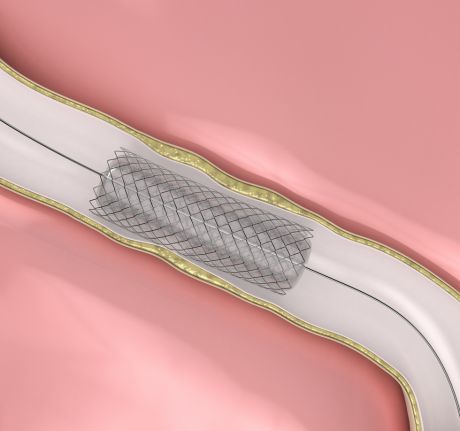
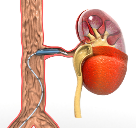
Technical Considerations for Stent Placement during URS

Selecting the Right Stent Size and Type

The first and most important technical aspect of stent placement is choosing the correct stent size and material. The stent needs to be long enough to cover the entire length of the ureter, ensuring it remains in place throughout the healing process. If the stent is too short or long, it may cause discomfort or fail to provide adequate support.

Factors to Consider When Choosing a Stent:

  • Ureteral Size and Anatomy: Ureter size and individual anatomical variations can affect the choice of stent. A smaller stent may be appropriate for a narrow ureter, while a larger one may be required for a wider ureter.
  • Material and Design: Most stents are made of silicone or polyurethane. Silicone stents are more flexible and are often preferred for their reduced likelihood of causing irritation. However, in cases of complex procedures or long-term stent placement, a polyurethane stent may be used.

Technical Tip: Dr. Anup Ramani, a urology oncology surgeon will consider the patient’s specific anatomical needs, stone location and overall health when selecting a stent for post-URS care.

What Our Patients Are Saying

"Dr Ramani is excellent not only as a best doctor but also a genuine and compassionate human being. He gives the kind of confidence with his immense experience over the years which is a life saver in a true sense. I say it from my personal experience that he is as good as it gets.its because of him it’s normal life and work."
Tanmoy Ganguli

Proper Insertion of the Stent

Once the correct stent has been selected, the next step is the insertion. A kidney stone ureteroscopy procedure typically involves using a guidewire to help insert the stent safely and precisely. It is essential that the stent is placed in the correct position to prevent migration or displacement, which can lead to infection or increased discomfort.

Technical Steps for Proper Stent Insertion:

  • Use of Guidewires: Guidewires help navigate the stent through the ureter and ensure it is correctly positioned without causing trauma to the tissue.
  • Secure Placement: The stent should be positioned with the lower end in the bladder and the upper end in the kidney, where it can prevent obstruction and allow urine to drain.

Technical Tip: Surgeons use real-time imaging to verify the placement of the stent, reducing the risk of complications and ensuring safety.

Managing Post-Procedure Discomfort

One of the biggest concerns with kidney stone ureteroscopy and stent placement is managing the post-procedure discomfort. After the procedure, the patient may experience pain or irritation due to the stent’s presence in the ureter. It is essential for the surgeon to anticipate these issues and provide the appropriate aftercare to minimize discomfort.

Post-Procedure Tips for Managing Discomfort:

  • Hydration: Increased fluid intake can help flush the urinary system, reducing irritation and helping the stent function more efficiently.
  • Stent Removal: While the stent is usually removed within a few weeks, it may cause ongoing discomfort until the removal process is complete. Discussing the stent removal timeline with the patient ensures they understand the process.

Technical Tip: Educating patients on what to expect post-procedure, including possible stent-related discomfort, is essential for improving their experience and managing anxiety.

Stent Removal and Follow-Up Care

After the kidney stone surgical treatment is complete, a follow-up procedure is typically required to remove the stent. This is usually done under local anesthesia and involves a simple cystoscopy procedure. Ensuring proper removal is critical to prevent complications like infection or the formation of stones around the stent.

Key Considerations for Stent Removal:

  • Timing of Removal: Stents are generally removed after 4-6 weeks, depending on the patient’s condition. Delaying stent removal can increase the risk of complications like encrustation or migration.
  • Monitoring for Complications: During the follow-up visit, the surgeon will check for any issues related to the stent or the healing of the ureter. If complications such as infection or pain persist, the doctor may recommend additional treatments.

Technical Tip: Proper timing of stent removal ensures optimal recovery and reduces the risk of complications. Surgeons use follow-up imaging and clinical evaluations to confirm the patient’s healing progress.

Contact Us for Kidney Stone Removal Surgery

Conclusion

The placement of a stent during kidney stone ureteroscopy is an essential step in ensuring successful recovery after kidney stone removal. By considering factors such as stent size, proper placement and managing post-operative discomfort, surgeons can help patients experience a smoother recovery. Stent-related complications, such as migration or infection, can be avoided with careful technical consideration and effective follow-up care.

For patients undergoing kidney stone removal surgery, particularly in India, working with an experienced Dr. Anup Ramani, a kidney stone surgeon ensures that all technical aspects of the procedure, including stent placement, are handled with precision and care. Whether for kidney stone removal by surgery, ensuring the right approach to stent placement can significantly improve patient safety, comfort and overall treatment outcomes.

FAQs

A stent is placed to ensure that the ureter remains open, preventing blockage and allowing for proper urine drainage during the healing process.

The stent is typically removed after 4-6 weeks, depending on the patient’s healing progress.

Yes, patients may experience discomfort, such as urinary urgency or pain during urination. Pain management and hydration are essential to reduce discomfort.

The stent is removed through a simple cystoscopy, a procedure done under local anesthesia.

Follow-up care includes monitoring the healing of the ureter, removal of the stent and ensuring that no complications such as infections or obstructions have occurred.

PARTIAL PENECTOMY
  • Partial penectomy is done in cases where glans and distal penis is involved with carcinoma. 
  • Partial penectomy is a type of organ-preserving surgery. Preservation of sexual and micturational function depends on the surgical dissection and reconstruction of residual urethra.
Kidney Stone Removal
  • Patients who develop stones in the kidney or ureter, often experience severe pain.
  • This condition usually needs a procedure to remove the kidney stones.
  • This procedure is called ureteroscopy and is performed very commonly.
  • It does not require any cuts and hence it is painless.
  • The procedure is performed with an endoscope inserted through the penis under spinal anesthesia.
  • The scope is inserted through the penis into the kidney and stones are dissolved with a laser.
  • The procedure takes about 40-50 minutes. 
  • A catheter (urine pipe) is kept after the procedure to drain the bladder. A stent is kept in the kidney at the same time.
  • Patient is mobile and walking in the room the same evening.
  • Hospital stay is one night and patient is discharged the next day after removal of the catheter.
  • Patient has to come back after six weeks to remove the stent in the kidney.
  • Patients can resume office a week after surgery and heavy activities like running, weight lifting, a month after the procedure.
  • We offer fixed packages for this procedure which can be obtained by calling our helpline +91 9967666060.
  • Men with an enlarged prostate, which is a normal ageing changes, often experiencing difficulty passing urine. This condition usually needs a procedure to trim the prostate and relieve the blockage.
  • This procedure is called TURP and is performed very commonly.
  • It does not require any cuts and hence it is painless.
  • The procedure is performed with an endoscope inserted through the penis under spinal anaesthesia.
  • The overgrown prostate is dissolved with a laser bloodlessly.
  • The procedure takes about 40 minutes.
  • A catheter (urine pipe) is kept after the procedure to drain the bladder.
  • Patient is mobile and walking in the room the same evening.
  • Hospital stay is two nights and patient is discharged with the catheter, which is removed after 4 days.
  • Patients can resume office a week after surgery and heavy activities like running, weight lifting, a month after the procedure.
  • We offer fixed packages for this procedure which can be obtained by calling our helpline +91 9967666060.
ROBOTIC ADRENALECTOMY FOR ADRENAL GLAND TUMOUR
  • Robotic adrenalectomy is a sophisticated, complex surgery and it is very important that an experienced surgeon performs this surgery to avoid major complications.

  • Once the anesthesia is done, and patient positioned, three micro cuts (3mm each) are made in the patient’s abdomen.

  • The arms of the Da Vinci robot are connected to the cuts via ports (tubes).

  • Dr. Ramani then sits in the controlling console to perform the surgery.

  • On an average, a robotic adrenalectomy takes one hour.

  • The surgery is almost completely bloodless and there has never been any need to transfuse blood after surgery.

  • A urine catheter and bag to drain the bladder is inserted during surgery.

  • A tiny drain pipe may be inserted in the surgical side of the abdomen, connected to a bag.

  • Patient is kept nil-by-mouth the day of the surgery, with IV fluids. Sips of water are started the next day and solid food by day three.

  • The drain pipe, if kept, is removed in the room on day 2 after surgery.

  • The catheter is removed on day two after surgery.

  • Total hospital stay for robotic adrenalectomy is 4 nights (including night before surgery).

  • Post discharge, a doctor from the surgical team visits the patient at home/ hotel room once every day.

RETURN TO ACTIVITY
  • On the day of discharge, patient is totally self-sufficient. They are able to walk freely without any pain, dress themselves, shower, toilet and they do not need to hire any nurse or help at home. Almost all patients are back to work within 2 weeks of surgery.

  • Heavy activities like running, weight lifting can be resumed after a month

FOLLOW UP AFTER SURGERY
  • Follow up after an adrenalectomy is in the form of CT scans, once a year for 5 years.
    Local patients usually meet Dr. Ramani after two weeks to discuss report.

  • Outstation patients are counselled on a phone consultation.

ROBOTIC SURGERY FOR BLADDER CANCER
  • Dr. Ramani is one of the very few surgeons in India who has the expertise to perform a robotic surgery for bladder cancer, which includes removing the urinary bladder and reconstructing a new bladder robotically.
  • Robotic radical cystectomy is an extremely sophisticated, complex surgery and it is very important that an experienced surgeon performs this surgery to avoid major complications.
  • Once the anaesthesia is done, and patient positioned, six micro cuts (3mm each) are made in the patient’s abdomen.
  • The arms of the Da Vinci robot are connected to the cuts via ports (tubes).
  • Dr. Ramani then sits in the controlling console to perform the surgery.
  • On an average, a robotic radical cystectomy with an ileal conduit takes 3-4 hours.
  • The surgery is almost completely bloodless and there has never been any need to transfuse blood after surgery.
  • A urine catheter and bag to drain the new bladder is inserted during surgery.
  • Two tiny drain pipe in inserted in the surgical side of the abdomen, connected to a bag.
  • Patient is kept nil-by-mouth for 4 days after surgery with IV supplementation of patient’s daily requirements of calories, fats, carbohydrates, proteins and electrolytes.
  • The drain pipes are removed in the room on day 3-5 after surgery.
  • Total hospital stay for radical cystectomy is 8 nights (including night before surgery).
  • Post discharge, a doctor from the surgical team visits the patient at home/ hotel room once every day.
RETURN TO ACTIVITY
  • On the day of discharge, patient is totally self-sufficient. They are able to walk freely without any pain, dress themselves, shower, toilet and they do not need to hire any nurse or help at home.
  • Almost all patients are back to work within 6 weeks of surgery. Heavy activities like running, weight lifting can be resumed after two months.
FOLLOW UP AFTER SURGERY
  • Follow up after a radical a cystectomy is in the form of CT scans, once a year for 5 years.

  • Histopathology report: Local patients usually meet Dr. Ramani after two weeks to discuss report.

  • Outstation patients are counselled on a phone consult. Depending on the report, patient may or may not need chemotherapy after surgery.

  • If chemo is needed, patients may choose to get it done with a medical oncologist of their choice or avail the services of one of the four medical oncologists on our team.

ROBOTIC RADICAL/PARTIAL NEPHRECTOMY FOR KIDNEY CANCER
  • Robotic partial nephrectomy is a sophisticated, complex surgery and it is very important that an experienced surgeon performs this surgery to avoid major complications. Robotic radical (total) nephrectomy is
  • relatively easier but still requires significant experience to consistently deliver results.
  • Once the anaesthesia is done, and patient positioned, five micro cuts (3mm each) are made in the patient’s abdomen.
  • The arms of the Da Vinci robot are connected to the cuts via ports (tubes).
  • Dr. Ramani then sits in the controlling console to perform the surgery.
  • On an average, a robotic radical nephrectomy takes one hour and a robotic partial nephrectomy takes about an hour and half.
  • The surgery is almost completely bloodless and there has never been any need to transfuse blood after surgery.
  • A urine catheter and bag to drain the bladder is inserted during surgery.
  • A tiny drain pipe in inserted in the surgical side of the abdomen, connected to a bag.
  • Patient is kept nil-by-mouth the day of the surgery, with IV fluids. Sips of water are started the next day and solid food by day three.
  • The drain pipe is removed in the room on day 3 after surgery. The catheter is removed on day two after surgery.
  • Total hospital stay for radical/partial nephrectomy is 4 nights (including night before surgery).
  • Post discharge, a doctor from the surgical team visits the patient at home/ hotel room once every day.
RETURN TO ACTIVITY
  • On the day of discharge, patient is totally self- sufficient. 
  • They are able to walk freely without any pain, dress themselves, shower, toilet and they do not need to hire any nurse or help at home. 
  • Almost all patients are back to work within 2-3 weeks of surgery.
  • Heavy activities like running, weight lifting can be resumed after a month.
FOLLOW UP AFTER SURGERY
  • Follow up after a radical/partial Nephrectomy is in the form of CT scans, once a year for 5 years.
  • Local patients usually meet Dr. Ramani after two weeks to discuss report. 
  • Outstation patients are counselled on a phone consultation.