




By Dr. Anup Ramani
Share At:
Google AI ChatGPT Grok Perplexity

Listening Article

AI Summary

When a kidney stone blocks the ureter, urine can no longer drain normally from the kidney to the bladder, and pressure builds up inside the kidney, often causing sudden, severe pain. What happens next depends on the stone’s size, its location, how severe the blockage is, kidney function, and the patient’s overall health. Small stones often pass on their own with pain control and hydration, while larger or more obstructive stones may need procedures such as ureteroscopy, laser stone removal, or shock wave lithotripsy. If the blockage causes infection or significant kidney strain, urgent drainage may be needed to protect kidney health.

Kidney Stone Blocking the Ureter: Introduction

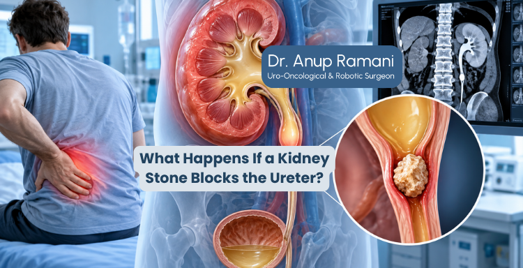
When a kidney stone blocks the ureter, it can cause urine flow obstruction, severe pain, and pressure buildup inside the kidney. The ureter is a narrow tube, only a few millimetres wide, that carries urine from each kidney down into the bladder, which is why even a small stone can get lodged inside it. A kidney stone blocking the ureter is one of the most common causes of sudden, intense flank pain, as the stone becomes stuck and urine backs up behind it.

Not every blocked ureter behaves the same way. Some patients pass the stone within days with minimal treatment, while others need active intervention, so every patient’s plan is different. A blocked ureter does not always mean permanent kidney damage. The risk depends on how long the blockage remains, whether infection is present, and how quickly urine flow is restored, and many patients recover fully once normal drainage returns.

 
Need More Information?
White Email Us Today!
 

What Happens When a Kidney Stone Blocks the Ureter?

Kidney stones form inside the kidney, and over time some dislodge and travel down into the ureter. A stone can get stuck at narrower points along the way, particularly where the ureter joins the kidney, crosses the pelvic blood vessels, or enters the bladder.

Once stuck, urine has nowhere to go. This causes:

  • Urine to accumulate above the blockage
  • Pressure to build up inside the kidney
  • Stretching of kidney tissue, which is often the source of pain

Doctors evaluate a blocked ureter using several factors together:

  • Stone size – smaller stones are more likely to pass on their own
  • Stone location – stones lower in the ureter pass more easily
  • Degree of blockage – partial obstruction behaves differently from complete blockage
  • Pain symptoms – severity often reflects how the kidney is being affected
  • Infection risk – a blocked, infected kidney is a medical emergency
  • Kidney function – existing kidney health affects treatment urgency

Blocked Ureter Symptoms: Signs of a Kidney Stone Blocking the Ureter

The classic symptom is sudden, severe pain in the side or back, often described as one of the worst pains a person can experience. It frequently comes in waves and may radiate towards the lower abdomen or groin as the stone moves.

Other common symptoms include:

  • Nausea and vomiting
  • Blood in the urine
  • Frequent urge to urinate
  • Burning sensation while urinating

These warning signs suggest a more serious situation needing urgent medical attention:

  • Fever and chills
  • Signs of a severe urinary infection
  • Noticeably reduced urine output

Hydronephrosis Due to Kidney Stone: How Ureter Blockage Affects Kidney Function

When urine flow is obstructed, pressure inside the kidney has nowhere to release, and the kidney’s collecting system begins to swell. This swelling, known as hydronephrosis due to kidney stone obstruction, is visible on imaging.

If the blockage is brief, the kidney usually recovers well once urine flow is restored. If it continues longer, pressure can affect the kidney’s filtering tissue and lead to a decline in kidney function, which is why doctors emphasise early diagnosis and ongoing kidney function monitoring.

Diagnosing a Kidney Stone Blocking the Ureter

Diagnosis usually begins with a medical history and physical examination focused on the pattern and location of pain, followed by:

  • Urine tests, to check for blood or infection
  • Blood tests, to assess kidney function
  • Ultrasound, a quick way to detect kidney swelling
  • CT scan, the most detailed imaging test for locating a stone precisely

CT scan is often preferred when doctors need detailed information about stone size, exact location, and degree of blockage.

These tests identify the stone’s size, location, and degree of obstruction, which guide the treatment decision that follows.

How Long Can a Kidney Stone Block the Ureter?

The length of time a blockage lasts plays a major role in how the kidney is affected.

  • A short blockage, lasting hours to a few days, often resolves without lasting kidney damage once the stone passes or is treated
  • A longer obstruction, extending over days to weeks, increases the risk of kidney function decline
  • If infection is present alongside the blockage, the situation becomes urgent regardless of how long the stone has been stuck

This is why doctors do not just look at the stone itself, but also at how long symptoms have been present when deciding how quickly to act.

Kidney Stone Treatment Options for a Blocked Ureter

Treatment selection depends on stone size, location, blockage severity, kidney function, and previous stone history, so the right approach is different for every patient.

Medicines and observation: Small stones, particularly those lower in the ureter, often have a reasonable chance of passing naturally with pain management, appropriate fluid intake, medication to relax the ureter, and close follow up.

Ureteroscopy and laser stone removal: For stones unlikely to pass on their own, a thin scope reaches the stone through the natural urinary passage, and laser technology breaks it into fragments that can then be removed or allowed to pass.

Shock wave lithotripsy: This non invasive technique uses focused shock waves from outside the body to break a stone into smaller pieces. It works best for certain stone sizes and locations, and recovery is generally straightforward.

Emergency procedures: When a blockage causes infection, severe pain, or kidney strain, urgent drainage may be needed first. This can involve a ureteral stent, a thin tube that allows urine to bypass the blockage, or nephrostomy drainage, a tube placed directly into the kidney through the skin, both restoring urine flow quickly to protect the kidney.

Kidney Stone Removal vs Natural Passage: Can a Blocked Ureter Heal Without Surgery?

Not every blocked ureter needs surgery.

  • Small stones may pass naturally with hydration and pain control
  • Not every blockage requires a procedure, especially when the stone is small and the kidney is not under significant strain
  • Procedures are generally considered when stones are large, persistent, causing infection, or affecting kidney function

Urine Flow Obstruction Due to Kidney Stone: When It Becomes an Emergency

Certain situations demand immediate attention: infection alongside blocked urine flow, worsening kidney function, pain that cannot be controlled, or complete obstruction of the ureter. In these situations, emergency drainage procedures may be necessary to relieve pressure and prevent further kidney damage, since delaying treatment can allow a treatable problem to become more serious.

Seek immediate medical attention if you have:

  • Fever with kidney stone pain
  • Chills
  • Very low or absent urine output
  • Severe, uncontrolled pain
  • A known history of kidney disease

What Our Patients Are Saying

" Dr. Ramani was very professional in his approach. He spent a lot of time to discuss the problem and compassionately handled a very complicated case. He is always accessible and ready to go the extra mile to put the patient at ease. Highly recommended! "
Manish Maheswari
On Google

Recovery After Kidney Stone Removal and Kidney Stone Recurrence Prevention

Recovery varies by procedure, but most patients return to normal activity within one to two weeks, with follow up appointments to confirm the stone has cleared and the kidney is recovering well. Good hydration and dietary adjustments based on the type of stone identified help support recovery and reduce the chance of recurrence.

Kidney stones tend to recur, so prevention is an important part of long term care. Adequate fluid intake, tailored dietary recommendations, and stone analysis when available all help a urologist build a personalised prevention strategy based on a patient’s stone history and diet.

A short period of obstruction is usually well tolerated by the kidney, but prolonged blockage can gradually affect kidney function, which is why timely treatment is emphasised so strongly. Monitoring kidney function after ureter blockage helps confirm recovery and identifies patients who may need closer long term follow up.

Modern minimally invasive procedures, such as ureteroscopy with laser stone removal, have made it possible to treat many kidney stones effectively with shorter recovery times compared to older surgical approaches. A thorough specialist evaluation, along with consistent follow up after kidney stone removal, helps determine the right approach and protects long term kidney health.

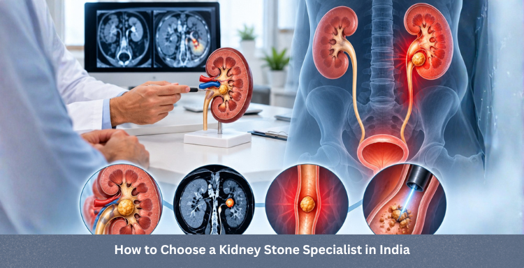
How to Choose a Kidney Stone Specialist in India

Important considerations include accurate evaluation of stone size, location, and severity of blockage, experience with a range of stone removal procedures, clear explanation of treatment options, a personalised approach to treatment planning, and a focus on long term kidney health rather than just immediate stone removal. An experienced urologist can help patients understand their options and make informed decisions about kidney stone management.

Dr. Anup Ramani: Kidney Stone Specialist in Mumbai

Dr. Anup Ramani is a urologist specialising in the diagnosis and treatment of urinary tract conditions, including kidney stones that cause ureter blockage, evaluating each case based on stone size, location, degree of blockage, and kidney function. As a kidney stone specialist in India offering kidney stone treatment in Mumbai, he focuses on personalised treatment planning, patient education, and evidence based care, including ureteroscopy and laser kidney stone removal. Patients experiencing severe kidney stone pain, urinary blockage symptoms, or repeated stones should consult an experienced urologist for timely evaluation and personalised treatment.

 
Call Now for Quick Support
White Call Now!
 

Conclusion

A kidney stone blocking the ureter can cause significant pain and urine flow obstruction, but most cases are manageable with timely diagnosis and the right treatment approach. Whether a stone passes naturally or requires a procedure depends on its size, location, and effect on kidney function. Consulting an experienced urologist early helps protect kidney health and prevent complications.

Kidney Stone Blocking the Ureter: Frequently Asked Questions

Urine flow from the kidney to the bladder is interrupted, causing pressure to build up inside the kidney, often with severe pain and, if untreated, a risk to kidney function.

Sudden, severe flank pain, blood in the urine, and nausea are common signs, confirmed with ultrasound or CT scan imaging.

Prolonged obstruction can affect kidney function, so timely diagnosis and treatment matter.

reatment ranges from pain management and observation for small stones to ureteroscopy, laser stone removal, or shock wave lithotripsy for larger or more obstructive stones.

Smaller stones, especially those lower in the ureter, often pass on their own with hydration and pain control, alongside medical monitoring.

When infection develops alongside blocked urine flow, kidney function is significantly affected, or pain cannot be controlled.

Most patients recover within one to two weeks, depending on the procedure and individual health factors.

Adequate hydration, dietary adjustments based on stone type, and regular urologist follow up reduce the risk of recurrence.

It is uncommon but possible, especially with a gradually forming or partial blockage. Silent blockages are sometimes found only on imaging done for other reasons, which is one reason regular checkups matter for patients with a history of stones.

No. Many small stones pass on their own with pain control and hydration. Surgery or a procedure is generally reserved for larger stones, stones that are not passing, or cases involving infection or significant kidney strain.

Untreated blockage can lead to worsening pain, infection, and progressive kidney damage. In severe or prolonged cases, it can permanently reduce how well the affected kidney functions, which is why timely evaluation is important.

In most cases, yes, especially when the blockage is identified and treated promptly. The kidney’s ability to recover depends largely on how long the obstruction lasted and whether infection was involved.

PARTIAL PENECTOMY
  • Partial penectomy is done in cases where glans and distal penis is involved with carcinoma. 
  • Partial penectomy is a type of organ-preserving surgery. Preservation of sexual and micturational function depends on the surgical dissection and reconstruction of residual urethra.
Kidney Stone Removal
  • Patients who develop stones in the kidney or ureter, often experience severe pain.
  • This condition usually needs a procedure to remove the kidney stones.
  • This procedure is called ureteroscopy and is performed very commonly.
  • It does not require any cuts and hence it is painless.
  • The procedure is performed with an endoscope inserted through the penis under spinal anesthesia.
  • The scope is inserted through the penis into the kidney and stones are dissolved with a laser.
  • The procedure takes about 40-50 minutes. 
  • A catheter (urine pipe) is kept after the procedure to drain the bladder. A stent is kept in the kidney at the same time.
  • Patient is mobile and walking in the room the same evening.
  • Hospital stay is one night and patient is discharged the next day after removal of the catheter.
  • Patient has to come back after six weeks to remove the stent in the kidney.
  • Patients can resume office a week after surgery and heavy activities like running, weight lifting, a month after the procedure.
  • We offer fixed packages for this procedure which can be obtained by calling our helpline +91 9967666060.
  • Men with an enlarged prostate, which is a normal ageing changes, often experiencing difficulty passing urine. This condition usually needs a procedure to trim the prostate and relieve the blockage.
  • This procedure is called TURP and is performed very commonly.
  • It does not require any cuts and hence it is painless.
  • The procedure is performed with an endoscope inserted through the penis under spinal anaesthesia.
  • The overgrown prostate is dissolved with a laser bloodlessly.
  • The procedure takes about 40 minutes.
  • A catheter (urine pipe) is kept after the procedure to drain the bladder.
  • Patient is mobile and walking in the room the same evening.
  • Hospital stay is two nights and patient is discharged with the catheter, which is removed after 4 days.
  • Patients can resume office a week after surgery and heavy activities like running, weight lifting, a month after the procedure.
  • We offer fixed packages for this procedure which can be obtained by calling our helpline +91 9967666060.
ROBOTIC ADRENALECTOMY FOR ADRENAL GLAND TUMOUR
  • Robotic adrenalectomy is a sophisticated, complex surgery and it is very important that an experienced surgeon performs this surgery to avoid major complications.

  • Once the anesthesia is done, and patient positioned, three micro cuts (3mm each) are made in the patient’s abdomen.

  • The arms of the Da Vinci robot are connected to the cuts via ports (tubes).

  • Dr. Ramani then sits in the controlling console to perform the surgery.

  • On an average, a robotic adrenalectomy takes one hour.

  • The surgery is almost completely bloodless and there has never been any need to transfuse blood after surgery.

  • A urine catheter and bag to drain the bladder is inserted during surgery.

  • A tiny drain pipe may be inserted in the surgical side of the abdomen, connected to a bag.

  • Patient is kept nil-by-mouth the day of the surgery, with IV fluids. Sips of water are started the next day and solid food by day three.

  • The drain pipe, if kept, is removed in the room on day 2 after surgery.

  • The catheter is removed on day two after surgery.

  • Total hospital stay for robotic adrenalectomy is 4 nights (including night before surgery).

  • Post discharge, a doctor from the surgical team visits the patient at home/ hotel room once every day.

RETURN TO ACTIVITY
  • On the day of discharge, patient is totally self-sufficient. They are able to walk freely without any pain, dress themselves, shower, toilet and they do not need to hire any nurse or help at home. Almost all patients are back to work within 2 weeks of surgery.

  • Heavy activities like running, weight lifting can be resumed after a month

FOLLOW UP AFTER SURGERY
  • Follow up after an adrenalectomy is in the form of CT scans, once a year for 5 years.
    Local patients usually meet Dr. Ramani after two weeks to discuss report.

  • Outstation patients are counselled on a phone consultation.

ROBOTIC SURGERY FOR BLADDER CANCER
  • Dr. Ramani is one of the very few surgeons in India who has the expertise to perform a robotic surgery for bladder cancer, which includes removing the urinary bladder and reconstructing a new bladder robotically.
  • Robotic radical cystectomy is an extremely sophisticated, complex surgery and it is very important that an experienced surgeon performs this surgery to avoid major complications.
  • Once the anaesthesia is done, and patient positioned, six micro cuts (3mm each) are made in the patient’s abdomen.
  • The arms of the Da Vinci robot are connected to the cuts via ports (tubes).
  • Dr. Ramani then sits in the controlling console to perform the surgery.
  • On an average, a robotic radical cystectomy with an ileal conduit takes 3-4 hours.
  • The surgery is almost completely bloodless and there has never been any need to transfuse blood after surgery.
  • A urine catheter and bag to drain the new bladder is inserted during surgery.
  • Two tiny drain pipe in inserted in the surgical side of the abdomen, connected to a bag.
  • Patient is kept nil-by-mouth for 4 days after surgery with IV supplementation of patient’s daily requirements of calories, fats, carbohydrates, proteins and electrolytes.
  • The drain pipes are removed in the room on day 3-5 after surgery.
  • Total hospital stay for radical cystectomy is 8 nights (including night before surgery).
  • Post discharge, a doctor from the surgical team visits the patient at home/ hotel room once every day.
RETURN TO ACTIVITY
  • On the day of discharge, patient is totally self-sufficient. They are able to walk freely without any pain, dress themselves, shower, toilet and they do not need to hire any nurse or help at home.
  • Almost all patients are back to work within 6 weeks of surgery. Heavy activities like running, weight lifting can be resumed after two months.
FOLLOW UP AFTER SURGERY
  • Follow up after a radical a cystectomy is in the form of CT scans, once a year for 5 years.

  • Histopathology report: Local patients usually meet Dr. Ramani after two weeks to discuss report.

  • Outstation patients are counselled on a phone consult. Depending on the report, patient may or may not need chemotherapy after surgery.

  • If chemo is needed, patients may choose to get it done with a medical oncologist of their choice or avail the services of one of the four medical oncologists on our team.

ROBOTIC RADICAL/PARTIAL NEPHRECTOMY FOR KIDNEY CANCER
  • Robotic partial nephrectomy is a sophisticated, complex surgery and it is very important that an experienced surgeon performs this surgery to avoid major complications. Robotic radical (total) nephrectomy is
  • relatively easier but still requires significant experience to consistently deliver results.
  • Once the anaesthesia is done, and patient positioned, five micro cuts (3mm each) are made in the patient’s abdomen.
  • The arms of the Da Vinci robot are connected to the cuts via ports (tubes).
  • Dr. Ramani then sits in the controlling console to perform the surgery.
  • On an average, a robotic radical nephrectomy takes one hour and a robotic partial nephrectomy takes about an hour and half.
  • The surgery is almost completely bloodless and there has never been any need to transfuse blood after surgery.
  • A urine catheter and bag to drain the bladder is inserted during surgery.
  • A tiny drain pipe in inserted in the surgical side of the abdomen, connected to a bag.
  • Patient is kept nil-by-mouth the day of the surgery, with IV fluids. Sips of water are started the next day and solid food by day three.
  • The drain pipe is removed in the room on day 3 after surgery. The catheter is removed on day two after surgery.
  • Total hospital stay for radical/partial nephrectomy is 4 nights (including night before surgery).
  • Post discharge, a doctor from the surgical team visits the patient at home/ hotel room once every day.
RETURN TO ACTIVITY
  • On the day of discharge, patient is totally self- sufficient. 
  • They are able to walk freely without any pain, dress themselves, shower, toilet and they do not need to hire any nurse or help at home. 
  • Almost all patients are back to work within 2-3 weeks of surgery.
  • Heavy activities like running, weight lifting can be resumed after a month.
FOLLOW UP AFTER SURGERY
  • Follow up after a radical/partial Nephrectomy is in the form of CT scans, once a year for 5 years.
  • Local patients usually meet Dr. Ramani after two weeks to discuss report. 
  • Outstation patients are counselled on a phone consultation.