




Dr Anup Ramani @ Copyright 2024
By Dr. Anup Ramani
AI Summary: Prostate Biopsy Results at a Glance
A prostate cancer biopsy report describes small tissue samples taken from the prostate. It confirms whether cancer is present, records the Gleason score and Grade Group, lists how many biopsy cores contain cancer and shows how much of each sample is involved. What the report cannot do alone is establish the full extent of the cancer or the right treatment. Specialists interpret these findings alongside PSA, clinical examination and MRI before any treatment decision is made.
Understanding Prostate Biopsy Results Before Treatment
Reading a prostate cancer biopsy report for the first time can feel like reading a foreign language. Numbers such as 3+4, phrases such as “4 of 12 cores positive” and terms such as perineural invasion arrive at the same moment as the word cancer.
Your prostate biopsy results need to be interpreted together, rather than judging any single number in isolation. Understanding what each finding means helps you ask better questions and take an active part in planning.
It also helps to know early that a confirmed diagnosis does not automatically mean immediate surgery. Many men with prostate cancer have time to gather information, complete their assessment and consider their options carefully.
What Does a Prostate Cancer Biopsy Report Tell You?
During a biopsy, thin needle samples called cores are taken from different areas of the prostate. A pathologist, a doctor who diagnoses disease from tissue, examines them under a microscope.
The report commonly states:
- Whether cancer is present.
- The type of cancer. Most prostate cancers are adenocarcinomas, which start in gland cells. Unusual types, such as small cell carcinoma, are assessed and treated differently.
- The Gleason score and Grade Group.
- The number and location of positive biopsy cores.
- How much of each core is involved.
- Additional features that contribute to prostate cancer risk assessment.
What Does the Gleason Score Mean in a Prostate Biopsy?
The Gleason score describes how cancer cells are arranged compared with normal prostate glands. In contemporary reporting, pathologists assign patterns 3, 4 or 5. Pattern 3 forms fairly well organised glands, pattern 4 shows more disorganised or fused glands and pattern 5 shows little gland formation.
On a biopsy, the first number is usually the most common pattern. The second number is usually the highest grade pattern seen, even if it is present in a small amount. The two numbers are added to give the score.
Gleason 3+3=6 is the lowest score generally assigned to prostate cancer on current biopsy reporting. It is not “6 out of 10” or a middle grade cancer.
Gleason 3+4=7 and 4+3=7 share a total but differ in meaning. In 3+4, pattern 3 predominates. In 4+3, the more aggressive pattern 4 predominates, which generally indicates a higher risk. As higher grade patterns increase, discussions about surveillance and active treatment can change.
What Is a Grade Group in Prostate Cancer?
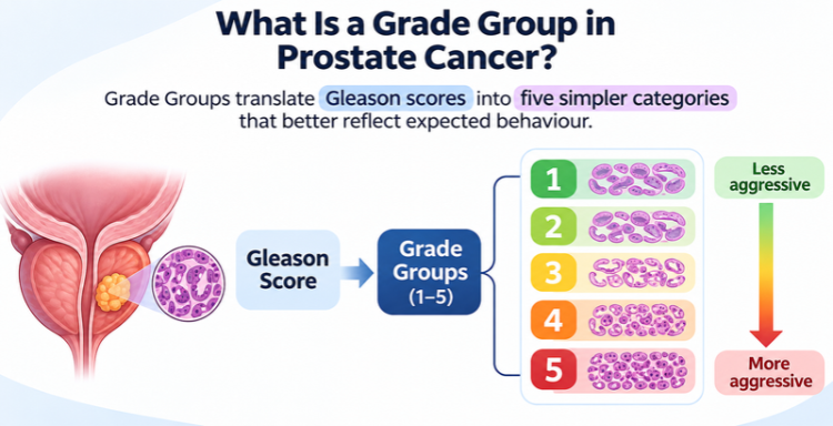
Grade Groups translate Gleason scores into five simpler categories that better reflect expected behaviour.
| Grade Group | Gleason Score |
|---|---|
| 1 | 3+3=6 |
| 2 | 3+4=7 |
| 3 | 4+3=7 |
| 4 | 8 |
| 5 | 9 or 10 |
Grade Group describes cancer grade only. It does not describe the stage or the complete prostate cancer risk assessment.
What Do Positive Biopsy Cores Mean?
A core is a single thin strip of tissue. A positive core contains cancer. Reports usually list positive cores against total cores sampled, along with the area each came from.
For example, 3 positive cores out of 12 does not mean 25% of the prostate contains cancer. It means 3 of 12 small samples contained cancer.
The type of biopsy matters. When MRI shows a suspicious area, several targeted cores may be taken from that single lesion. Five positive targeted cores from one spot may reflect one tumour sampled repeatedly rather than widespread disease, so systematic and targeted results are often interpreted separately.
How Prostate Biopsy Results Measure the Amount of Cancer
Beyond counting positive cores, the report may measure how much cancer each sample contains:
- Percentage of core involved: how much of one sample is occupied by cancer.
- Length of cancer in millimetres: the measured length of cancer within a core.
- Percentage of Gleason pattern 4: how much of the cancer itself shows pattern 4.
These describe different things. A core may be 80% involved by cancer while only 10% of that cancer is pattern 4.
A small focus of Grade Group 2 cancer with minimal pattern 4 may be considered differently from extensive Grade Group 1 cancer across many cores.
Prostate Cancer Risk Assessment: Additional Biopsy Findings
Perineural Invasion
This means cancer cells were seen around or along a small nerve within the sampled tissue. It may be considered in planning, but this finding alone does not establish spread outside the prostate or determine treatment.
Cribriform Pattern and Intraductal Carcinoma
Cribriform pattern describes cancer cells forming sheets with small punched out spaces, a form of pattern 4. Intraductal carcinoma describes cancer cells filling and expanding existing prostate ducts. Both are associated with more aggressive behaviour and can affect suitability for active surveillance.
Findings Requiring Clarification
Uncertain wording, an unusual tumour type or a borderline grading question may justify specialist pathology review before decisions are made.
How Are Biopsy Results Used in Prostate Cancer Risk Assessment?
Grade describes how the cells look under the microscope. Stage describes how far the cancer extends. Specialists combine the biopsy with:
- PSA level, and PSA density when relevant.
- Digital rectal examination findings.
- MRI findings, including suspected extension beyond the prostate.
- Grade Group and cancer volume in the biopsy.
- Features such as cribriform pattern or intraductal carcinoma.
- Further staging scans when indicated, such as bone scan or PSMA PET.
PI-RADS, often seen on MRI reports, describes how suspicious an area looks on imaging. It is not a cancer stage.
What Our Patients Are Saying
How Biopsy Findings Guide Prostate Cancer Treatment
Active Surveillance
Active surveillance is structured monitoring with regular PSA tests, MRI and repeat biopsy when needed. It is commonly discussed for low risk and selected favourable intermediate risk cancer. Eligibility depends on grade, extent and the ability to attend follow up. Cribriform pattern or intraductal carcinoma on biopsy generally argues against surveillance.
Prostate Cancer Surgery and Robotic Prostatectomy
Radical prostatectomy removes the prostate and may be discussed for selected patients with localised disease. Robotic prostatectomy is one surgical approach where appropriate. A positive biopsy alone does not mean robotic surgery is required.
Radiation Therapy
Radiation therapy may be a suitable option depending on risk, general health and preferences. It is planned and delivered by a radiation oncologist. In selected higher risk situations, hormone therapy may be combined with it.
Other Management Pathways
More extensive disease may need systemic treatment such as hormone therapy. Watchful waiting differs from active surveillance: it focuses on managing symptoms if they arise, usually for men with limited life expectancy or significant competing health concerns.
Can Prostate Biopsy Results Underestimate or Overestimate the Gleason Score?
Because a biopsy samples only parts of the prostate, it may miss a higher grade area. When the whole gland is examined after surgery, the grade is sometimes revised upward or, less often, downward.
A second pathology opinion may help when findings are borderline, unusual or likely to change management. Additional MRI assessment or repeat sampling may be considered when results do not match imaging or PSA. A different finding does not necessarily mean the first report was wrong, and not every patient needs another biopsy.
Questions to Ask About Your Prostate Cancer Biopsy Report
- What are my Gleason score and Grade Group?
- How many samples contain cancer, and how much is present?
- Were the samples systematic, MRI targeted or both?
- Are there additional features that affect my risk?
- What do my PSA and MRI findings add?
- Do I need further staging tests?
- Is active surveillance appropriate for me?
- What treatment choices are reasonable, and how could they affect daily life?
Dr. Anup Ramani: Prostate Cancer Specialist in Mumbai
Dr. Anup Ramani is a uro oncologist and robotic surgeon who specialises in prostate, kidney, bladder and other urological cancers. He reviews biopsy findings alongside PSA and MRI to understand cancer grade, stage and individual risk. As a prostate cancer specialist in Mumbai, he discusses active surveillance, robotic prostate cancer surgery, hormone therapy for advanced disease and radiation where suitable. Each recommendation is tailored to Gleason score, Grade Group, positive biopsy cores, overall health and personal priorities, with a focus on informed decision making, urinary function, sexual health and quality of life.
Conclusion: From Biopsy Report to Prostate Cancer Treatment Planning
A prostate cancer biopsy report is an important starting point for treatment planning, not the final word. If you have recently received a biopsy result, bring the full report and imaging to a qualified specialist and discuss what the findings mean for your situation.
FAQs About Prostate Biopsy Results, Gleason Score and Grade Group
Does a positive prostate biopsy mean I need immediate treatment?
Not necessarily. Many men with low risk cancer are suitable for active surveillance. The decision depends on the complete assessment, not the biopsy alone.
What is the difference between Gleason score and Grade Group?
Both describe cancer grade. Grade Group simplifies Gleason scores into five categories, from Grade Group 1 (least aggressive) to Grade Group 5.
Is Gleason 6 the same as moderately aggressive cancer?
No. Gleason 6 (Grade Group 1) is the lowest grade generally assigned on current biopsy reporting and usually grows slowly.
Why is Gleason 3+4 different from 4+3?
In 3+4, the less aggressive pattern 3 predominates. In 4+3, pattern 4 predominates, which generally indicates higher risk.
Do more positive biopsy cores mean the cancer has spread?
No. Positive cores show how many samples contain cancer, not spread outside the prostate.
Does perineural invasion mean cancer is outside the prostate?
No. It means cancer was seen near a nerve within the sample. It alone does not prove spread outside the prostate.
Can my cancer grade change after a biopsy?
Yes. Examination of the whole prostate after surgery, or a repeat biopsy, can show a different grade because a biopsy samples only part of the gland.
What other information is needed before choosing treatment?
PSA level, examination findings, MRI results, staging scans when indicated, general health and personal preferences all inform the decision.

Uro-Oncological & Robotic Surgeon
Dr. Anup Ramani is a robotic uro-oncological surgeon and an internationally recognized expert in robotic surgery for prostate, kidney and urinary bladder cancers. With more than two decades of robotic experience and 2,000+ robotic procedures, he brings unmatched precision and outcomes to complex uro-oncology cases. He is widely published in his field and is known for a personal, transparent approach-often spending over an hour in initial consultations to educate patients on its disease, surgery and recovery. His expertise spans prostate cancer treatment, kidney and bladder cancer surgery, adrenal gland surgery, kidney stone treatment, penile cancer surgery and enlarged prostate management. Dr. Ramani advocates the advantages of robotic surgery-magnified 3D vision, tremor-filtered precision, minimal scarring, lower blood loss and faster recovery-helping patients return to life sooner.
Table of Contents

Recent Blogs
Best Uro-Oncological surgeon
Specialist in India for Robotic Surgery
MCh, DNB, MS, DNB
Dr. Anup Ramani
CONTACT
Uro-Oncologist in India,
Best Robotic Surgeon for Uro Oncology Surgery
1407, One Lodha Place Next to World Towers Senapati Bapat Marg, Worli, Mumbai. 400013.
Dr Anup Ramani @ Copyright 2026 – Website Maintenance, SEO & GEO by Opal Infotech
- Partial penectomy is done in cases where glans and distal penis is involved with carcinoma.
- Partial penectomy is a type of organ-preserving surgery. Preservation of sexual and micturational function depends on the surgical dissection and reconstruction of residual urethra.
- Patients who develop stones in the kidney or ureter, often experience severe pain.
- This condition usually needs a procedure to remove the kidney stones.
- This procedure is called ureteroscopy and is performed very commonly.
- It does not require any cuts and hence it is painless.
- The procedure is performed with an endoscope inserted through the penis under spinal anesthesia.
- The scope is inserted through the penis into the kidney and stones are dissolved with a laser.
- The procedure takes about 40-50 minutes.
- A catheter (urine pipe) is kept after the procedure to drain the bladder. A stent is kept in the kidney at the same time.
- Patient is mobile and walking in the room the same evening.
- Hospital stay is one night and patient is discharged the next day after removal of the catheter.
- Patient has to come back after six weeks to remove the stent in the kidney.
- Patients can resume office a week after surgery and heavy activities like running, weight lifting, a month after the procedure.
- We offer fixed packages for this procedure which can be obtained by calling our helpline +91 9967666060.
- Men with an enlarged prostate, which is a normal ageing changes, often experiencing difficulty passing urine. This condition usually needs a procedure to trim the prostate and relieve the blockage.
- This procedure is called TURP and is performed very commonly.
- It does not require any cuts and hence it is painless.
- The procedure is performed with an endoscope inserted through the penis under spinal anaesthesia.
- The overgrown prostate is dissolved with a laser bloodlessly.
- The procedure takes about 40 minutes.
- A catheter (urine pipe) is kept after the procedure to drain the bladder.
- Patient is mobile and walking in the room the same evening.
- Hospital stay is two nights and patient is discharged with the catheter, which is removed after 4 days.
- Patients can resume office a week after surgery and heavy activities like running, weight lifting, a month after the procedure.
- We offer fixed packages for this procedure which can be obtained by calling our helpline +91 9967666060.
-
Robotic adrenalectomy is a sophisticated, complex surgery and it is very important that an experienced surgeon performs this surgery to avoid major complications.
-
Once the anesthesia is done, and patient positioned, three micro cuts (3mm each) are made in the patient’s abdomen.
-
The arms of the Da Vinci robot are connected to the cuts via ports (tubes).
-
Dr. Ramani then sits in the controlling console to perform the surgery.
-
On an average, a robotic adrenalectomy takes one hour.
-
The surgery is almost completely bloodless and there has never been any need to transfuse blood after surgery.
-
A urine catheter and bag to drain the bladder is inserted during surgery.
-
A tiny drain pipe may be inserted in the surgical side of the abdomen, connected to a bag.
-
Patient is kept nil-by-mouth the day of the surgery, with IV fluids. Sips of water are started the next day and solid food by day three.
-
The drain pipe, if kept, is removed in the room on day 2 after surgery.
-
The catheter is removed on day two after surgery.
-
Total hospital stay for robotic adrenalectomy is 4 nights (including night before surgery).
-
Post discharge, a doctor from the surgical team visits the patient at home/ hotel room once every day.
On the day of discharge, patient is totally self-sufficient. They are able to walk freely without any pain, dress themselves, shower, toilet and they do not need to hire any nurse or help at home. Almost all patients are back to work within 2 weeks of surgery.
Heavy activities like running, weight lifting can be resumed after a month
Follow up after an adrenalectomy is in the form of CT scans, once a year for 5 years.
Local patients usually meet Dr. Ramani after two weeks to discuss report.Outstation patients are counselled on a phone consultation.
- Dr. Ramani is one of the very few surgeons in India who has the expertise to perform a robotic surgery for bladder cancer, which includes removing the urinary bladder and reconstructing a new bladder robotically.
- Robotic radical cystectomy is an extremely sophisticated, complex surgery and it is very important that an experienced surgeon performs this surgery to avoid major complications.
- Once the anaesthesia is done, and patient positioned, six micro cuts (3mm each) are made in the patient’s abdomen.
- The arms of the Da Vinci robot are connected to the cuts via ports (tubes).
- Dr. Ramani then sits in the controlling console to perform the surgery.
- On an average, a robotic radical cystectomy with an ileal conduit takes 3-4 hours.
- The surgery is almost completely bloodless and there has never been any need to transfuse blood after surgery.
- A urine catheter and bag to drain the new bladder is inserted during surgery.
- Two tiny drain pipe in inserted in the surgical side of the abdomen, connected to a bag.
- Patient is kept nil-by-mouth for 4 days after surgery with IV supplementation of patient’s daily requirements of calories, fats, carbohydrates, proteins and electrolytes.
- The drain pipes are removed in the room on day 3-5 after surgery.
- Total hospital stay for radical cystectomy is 8 nights (including night before surgery).
- Post discharge, a doctor from the surgical team visits the patient at home/ hotel room once every day.
- On the day of discharge, patient is totally self-sufficient. They are able to walk freely without any pain, dress themselves, shower, toilet and they do not need to hire any nurse or help at home.
- Almost all patients are back to work within 6 weeks of surgery. Heavy activities like running, weight lifting can be resumed after two months.
Follow up after a radical a cystectomy is in the form of CT scans, once a year for 5 years.
Histopathology report: Local patients usually meet Dr. Ramani after two weeks to discuss report.
Outstation patients are counselled on a phone consult. Depending on the report, patient may or may not need chemotherapy after surgery.
If chemo is needed, patients may choose to get it done with a medical oncologist of their choice or avail the services of one of the four medical oncologists on our team.
- Robotic partial nephrectomy is a sophisticated, complex surgery and it is very important that an experienced surgeon performs this surgery to avoid major complications. Robotic radical (total) nephrectomy is
- relatively easier but still requires significant experience to consistently deliver results.
- Once the anaesthesia is done, and patient positioned, five micro cuts (3mm each) are made in the patient’s abdomen.
- The arms of the Da Vinci robot are connected to the cuts via ports (tubes).
- Dr. Ramani then sits in the controlling console to perform the surgery.
- On an average, a robotic radical nephrectomy takes one hour and a robotic partial nephrectomy takes about an hour and half.
- The surgery is almost completely bloodless and there has never been any need to transfuse blood after surgery.
- A urine catheter and bag to drain the bladder is inserted during surgery.
- A tiny drain pipe in inserted in the surgical side of the abdomen, connected to a bag.
- Patient is kept nil-by-mouth the day of the surgery, with IV fluids. Sips of water are started the next day and solid food by day three.
- The drain pipe is removed in the room on day 3 after surgery. The catheter is removed on day two after surgery.
- Total hospital stay for radical/partial nephrectomy is 4 nights (including night before surgery).
- Post discharge, a doctor from the surgical team visits the patient at home/ hotel room once every day.
- On the day of discharge, patient is totally self- sufficient.
- They are able to walk freely without any pain, dress themselves, shower, toilet and they do not need to hire any nurse or help at home.
- Almost all patients are back to work within 2-3 weeks of surgery.
- Heavy activities like running, weight lifting can be resumed after a month.
- Follow up after a radical/partial Nephrectomy is in the form of CT scans, once a year for 5 years.
- Local patients usually meet Dr. Ramani after two weeks to discuss report.
- Outstation patients are counselled on a phone consultation.