Dr Anup Ramani @ Copyright 2024
By Dr. Anup Ramani
Kidney stones, also known as renal calculi, are solid masses made of minerals and salts that form inside the kidneys. These stones can vary in size from being as small as a grain of sand to as large as a golf ball. While they may pass through the urinary system without causing significant issues, larger stones can lead to severe pain and require medical intervention. Understanding the causes, types, and early warning signs of kidney stones can help individuals prevent or manage the condition effectively.
What Are Kidney Stones?
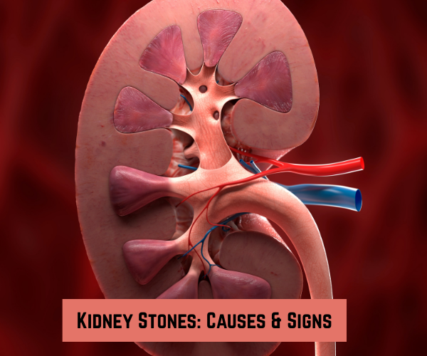
Kidney stones are hardened deposits of minerals and salts that form in the kidneys. They develop when the urine becomes highly concentrated, allowing minerals and salts to crystallize and clump together. These stones can form in the kidneys and travel through the urinary tract as they grow in size.
While some stones may pass without causing symptoms, larger ones can block the ureter, causing pain, bleeding, and other complications.
What Our Patients Are Saying
What Are the Causes of Kidney Stones?
Several factors can contribute to the formation of kidney stones. Here are the primary causes:
Dehydration
- Not drinking enough fluids can lead to concentrated urine, which makes it easier for minerals and salts to crystallize and form stones. Adequate hydration helps dilute the substances in the urine, reducing the risk of stone formation.
Dietary Factors
- High sodium intake: Excessive salt can increase calcium levels in the urine, which raises the risk of forming calcium-based stones.
- High animal protein diet: Diets rich in beef, fish, and chicken can make the urine more acidic, which increases the risk of forming uric acid stones.
- Oxalate-rich foods: Foods like spinach, chocolate, and nuts contain high levels of oxalates, which can combine with calcium in the kidneys to form calcium oxalate stones.
Family and Personal History
- A family history of kidney stones significantly increases the chances of developing them. Individuals who have previously had kidney stones are also at higher risk of recurrence.
Medical Conditions
Certain health conditions can increase the likelihood of kidney stone formation:

- Hyperparathyroidism: This condition leads to excess calcium in the blood, which can be filtered into the kidneys, forming calcium stones.
- Gout: A disorder that causes high levels of uric acid in the blood, contributing to the formation of uric acid stones.
- Diabetes and Obesity: Both conditions can increase the risk of kidney stones due to changes in urine composition and metabolic factors.
- Urinary Tract Infections (UTIs): UTIs can increase the risk of forming struvite stones, a type of stone that forms due to bacterial infections.
Medications
Some medications and supplements, including vitamin C and calcium supplements, may increase the risk of kidney stones. Overuse of diuretics or certain antacids can also contribute to stone formation.
What Are the Different Types of Kidney Stones?
There are four primary types of kidney stones, each with different causes and risk factors:
Calcium Stones
- Most common type of kidney stones, typically composed of calcium oxalate or calcium phosphate.
- These stones can form when there is too much calcium or oxalate in the urine, often due to diet, dehydration, or an underlying medical condition like hyperparathyroidism.
Uric Acid Stones
- These form when the urine becomes too acidic, often as a result of a high-protein diet (especially red meat), dehydration, or conditions like gout.
- Uric acid stones can also be associated with metabolic disorders and certain cancers.
Struvite Stones
- These stones are less common and typically occur as a result of urinary tract infections (UTIs).
- Struvite stones are often larger and can grow quickly, potentially causing obstruction in the urinary tract.
Cystine Stones
- Rare type of kidney stones that form due to a genetic disorder called cystinuria, where the kidneys leak excess cystine (an amino acid) into the urine.
- People with cystinuria produce stones much earlier in life compared to others.
What Are the Early Warning Signs of Kidney Stones?
Kidney stones often don’t cause symptoms until they move into the ureter, the tube that connects the kidney to the bladder. However, recognizing the early signs can help with prompt diagnosis and treatment. Common warning signs include:
Severe Pain (Renal Colic)
- Often described as sharp, stabbing, or cramp-like pain in the back, side, or lower abdomen.
- The pain may radiate to the groin area and can fluctuate in intensity, coming in waves as the stone moves through the urinary tract.
Painful or Frequent Urination
- Burning or painful urination can occur as the stone irritates the urinary tract.
- Increased frequency of urination or a feeling of urgency to urinate may also accompany kidney stones.
Blood in the Urine (Hematuria)
- The presence of blood in the urine, which can cause it to appear pink, red, or brown, is a common sign of kidney stones.
- Blood occurs when the stone irritates the urinary tract lining, causing bleeding.
Cloudy or Foul-Smelling Urine
- Cloudy or foul-smelling urine may indicate a urinary tract infection (UTI), which can sometimes accompany kidney stones.
- UTIs can lead to the formation of struvite stones and worsen existing kidney stones.
Nausea and Vomiting
- These symptoms may be caused by the severe pain associated with kidney stones or the body’s response to the stone in the urinary tract.
Difficulty Passing Urine
- A blockage in the urinary tract caused by a stone can slow down or stop the flow of urine.
- This can cause discomfort and may require immediate medical attention to avoid further complications.
Fever and Chills
- If a kidney stone causes an infection, it can lead to fever and chills.
- These are signs of a serious infection and require immediate medical attention to prevent complications like sepsis.
What Should You Do If You Experience Symptoms of Kidney Stones?
If you suspect you have kidney stones or experience any of the severe symptoms, especially fever or difficulty urinating, it’s crucial to seek medical attention immediately. A healthcare provider will diagnose the stones through:
- Imaging tests like CT scans or ultrasound.
- Urine tests to detect blood or crystals.
- Blood tests to check for calcium, uric acid, or infection.
Prompt diagnosis and treatment can help prevent serious complications like kidney damage or urinary tract infections.
How Can Kidney Stones Be Prevented?
While not all kidney stones are preventable, there are several steps you can take to reduce your risk:
- Stay hydrated: Drink plenty of water to dilute your urine and reduce the likelihood of stone formation.
- Limit sodium intake: Avoid high-sodium foods to prevent calcium from accumulating in the urine.
- Reduce animal protein consumption: Lower intake of red meat, poultry, and fish, as these can increase the risk of uric acid stones.
- Avoid oxalate-rich foods: Limit foods high in oxalates, such as spinach, nuts, and chocolate, especially if you have a history of calcium oxalate stones.
Maintain a healthy weight: Obesity is a known risk factor for kidney stones, so regular exercise and a balanced diet are essential.
Conclusion
Kidney stones are a common yet often painful condition that affects many people worldwide. Understanding the causes, types, and early warning signs of kidney stones can help with early detection and treatment. If you experience symptoms like severe pain, blood in the urine, or difficulty urinating, it’s important to seek medical help immediately to avoid complications. While some stones may be preventable through lifestyle modifications, early diagnosis and proper care are crucial for managing kidney stones and improving overall health.
Table of Contents
Recent Blogs
Best Uro-Oncological surgeon
Specialist in India for Robotic Surgery
MCh, DNB, MS, DNB
Dr. Anup Ramani
CONTACT
Uro-Oncologist in India,
Best Robotic Surgeon for Uro Oncology Surgery
1407, One Lodha Place Next to World Towers Senapati Bapat Marg, Worli, Mumbai. 400013.
Dr Anup Ramani @ Copyright 2024 – Website Maintenance & SEO by Webmasterindia
- Partial penectomy is done in cases where glans and distal penis is involved with carcinoma.
- Partial penectomy is a type of organ-preserving surgery. Preservation of sexual and micturational function depends on the surgical dissection and reconstruction of residual urethra.
- Patients who develop stones in the kidney or ureter, often experience severe pain.
- This condition usually needs a procedure to remove the kidney stones.
- This procedure is called ureteroscopy and is performed very commonly.
- It does not require any cuts and hence it is painless.
- The procedure is performed with an endoscope inserted through the penis under spinal anesthesia.
- The scope is inserted through the penis into the kidney and stones are dissolved with a laser.
- The procedure takes about 40-50 minutes.
- A catheter (urine pipe) is kept after the procedure to drain the bladder. A stent is kept in the kidney at the same time.
- Patient is mobile and walking in the room the same evening.
- Hospital stay is one night and patient is discharged the next day after removal of the catheter.
- Patient has to come back after six weeks to remove the stent in the kidney.
- Patients can resume office a week after surgery and heavy activities like running, weight lifting, a month after the procedure.
- We offer fixed packages for this procedure which can be obtained by calling our helpline +91 9967666060.
- Men with an enlarged prostate, which is a normal ageing changes, often experiencing difficulty passing urine. This condition usually needs a procedure to trim the prostate and relieve the blockage.
- This procedure is called TURP and is performed very commonly.
- It does not require any cuts and hence it is painless.
- The procedure is performed with an endoscope inserted through the penis under spinal anaesthesia.
- The overgrown prostate is dissolved with a laser bloodlessly.
- The procedure takes about 40 minutes.
- A catheter (urine pipe) is kept after the procedure to drain the bladder.
- Patient is mobile and walking in the room the same evening.
- Hospital stay is two nights and patient is discharged with the catheter, which is removed after 4 days.
- Patients can resume office a week after surgery and heavy activities like running, weight lifting, a month after the procedure.
- We offer fixed packages for this procedure which can be obtained by calling our helpline +91 9967666060.
-
Robotic adrenalectomy is a sophisticated, complex surgery and it is very important that an experienced surgeon performs this surgery to avoid major complications.
-
Once the anesthesia is done, and patient positioned, three micro cuts (3mm each) are made in the patient’s abdomen.
-
The arms of the Da Vinci robot are connected to the cuts via ports (tubes).
-
Dr. Ramani then sits in the controlling console to perform the surgery.
-
On an average, a robotic adrenalectomy takes one hour.
-
The surgery is almost completely bloodless and there has never been any need to transfuse blood after surgery.
-
A urine catheter and bag to drain the bladder is inserted during surgery.
-
A tiny drain pipe may be inserted in the surgical side of the abdomen, connected to a bag.
-
Patient is kept nil-by-mouth the day of the surgery, with IV fluids. Sips of water are started the next day and solid food by day three.
-
The drain pipe, if kept, is removed in the room on day 2 after surgery.
-
The catheter is removed on day two after surgery.
-
Total hospital stay for robotic adrenalectomy is 4 nights (including night before surgery).
-
Post discharge, a doctor from the surgical team visits the patient at home/ hotel room once every day.
On the day of discharge, patient is totally self-sufficient. They are able to walk freely without any pain, dress themselves, shower, toilet and they do not need to hire any nurse or help at home. Almost all patients are back to work within 2 weeks of surgery.
Heavy activities like running, weight lifting can be resumed after a month
Follow up after an adrenalectomy is in the form of CT scans, once a year for 5 years.
Local patients usually meet Dr. Ramani after two weeks to discuss report.Outstation patients are counselled on a phone consultation.
- Dr. Ramani is one of the very few surgeons in India who has the expertise to perform a robotic surgery for bladder cancer, which includes removing the urinary bladder and reconstructing a new bladder robotically.
- Robotic radical cystectomy is an extremely sophisticated, complex surgery and it is very important that an experienced surgeon performs this surgery to avoid major complications.
- Once the anaesthesia is done, and patient positioned, six micro cuts (3mm each) are made in the patient’s abdomen.
- The arms of the Da Vinci robot are connected to the cuts via ports (tubes).
- Dr. Ramani then sits in the controlling console to perform the surgery.
- On an average, a robotic radical cystectomy with an ileal conduit takes 3-4 hours.
- The surgery is almost completely bloodless and there has never been any need to transfuse blood after surgery.
- A urine catheter and bag to drain the new bladder is inserted during surgery.
- Two tiny drain pipe in inserted in the surgical side of the abdomen, connected to a bag.
- Patient is kept nil-by-mouth for 4 days after surgery with IV supplementation of patient’s daily requirements of calories, fats, carbohydrates, proteins and electrolytes.
- The drain pipes are removed in the room on day 3-5 after surgery.
- Total hospital stay for radical cystectomy is 8 nights (including night before surgery).
- Post discharge, a doctor from the surgical team visits the patient at home/ hotel room once every day.
- On the day of discharge, patient is totally self-sufficient. They are able to walk freely without any pain, dress themselves, shower, toilet and they do not need to hire any nurse or help at home.
- Almost all patients are back to work within 6 weeks of surgery. Heavy activities like running, weight lifting can be resumed after two months.
Follow up after a radical a cystectomy is in the form of CT scans, once a year for 5 years.
Histopathology report: Local patients usually meet Dr. Ramani after two weeks to discuss report.
Outstation patients are counselled on a phone consult. Depending on the report, patient may or may not need chemotherapy after surgery.
If chemo is needed, patients may choose to get it done with a medical oncologist of their choice or avail the services of one of the four medical oncologists on our team.
- Robotic partial nephrectomy is a sophisticated, complex surgery and it is very important that an experienced surgeon performs this surgery to avoid major complications. Robotic radical (total) nephrectomy is
- relatively easier but still requires significant experience to consistently deliver results.
- Once the anaesthesia is done, and patient positioned, five micro cuts (3mm each) are made in the patient’s abdomen.
- The arms of the Da Vinci robot are connected to the cuts via ports (tubes).
- Dr. Ramani then sits in the controlling console to perform the surgery.
- On an average, a robotic radical nephrectomy takes one hour and a robotic partial nephrectomy takes about an hour and half.
- The surgery is almost completely bloodless and there has never been any need to transfuse blood after surgery.
- A urine catheter and bag to drain the bladder is inserted during surgery.
- A tiny drain pipe in inserted in the surgical side of the abdomen, connected to a bag.
- Patient is kept nil-by-mouth the day of the surgery, with IV fluids. Sips of water are started the next day and solid food by day three.
- The drain pipe is removed in the room on day 3 after surgery. The catheter is removed on day two after surgery.
- Total hospital stay for radical/partial nephrectomy is 4 nights (including night before surgery).
- Post discharge, a doctor from the surgical team visits the patient at home/ hotel room once every day.
- On the day of discharge, patient is totally self- sufficient.
- They are able to walk freely without any pain, dress themselves, shower, toilet and they do not need to hire any nurse or help at home.
- Almost all patients are back to work within 2-3 weeks of surgery.
- Heavy activities like running, weight lifting can be resumed after a month.
- Follow up after a radical/partial Nephrectomy is in the form of CT scans, once a year for 5 years.
- Local patients usually meet Dr. Ramani after two weeks to discuss report.
- Outstation patients are counselled on a phone consultation.