




By Dr. Anup Ramani
Share At:
Google AI ChatGPT Grok Perplexity

Listening Article

AI Summary:

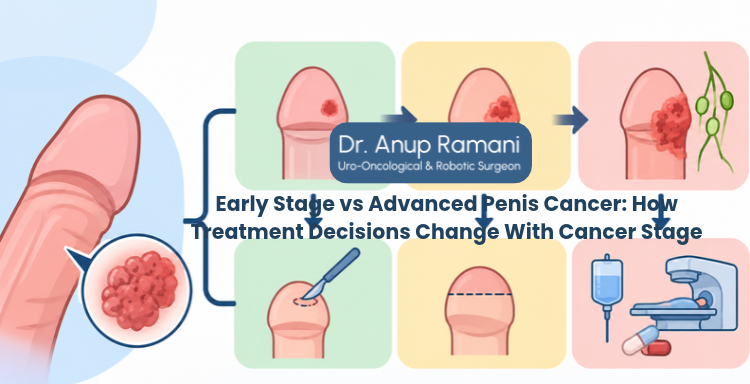
Penis cancer stages and treatment are closely linked. Treatment depends on how deeply the tumour has grown, whether groin or pelvic lymph nodes are involved and whether cancer has reached distant organs. Selected superficial or small localised tumours may be treated with approaches that preserve the penis, while deeper tumours or lymph node disease may need more extensive surgery or combined treatment. Advanced cancer does not always mean distant spread, and every plan needs individual assessment.

Why Penis Cancer Treatment Plans Differ

Two men with the same diagnosis can receive very different treatment. Penis cancer and penile cancer are the same condition, and most cases are squamous cell carcinomas; rarer types are managed differently.

Early stage penis cancer treatment may preserve healthy tissue, while penile cancer surgery options range from local excision to partial or total penectomy. Advanced penile cancer treatment may add lymph node surgery, chemotherapy or radiation.

 
We’re Ready to Help!
White Email Us
 

Penile Cancer Staging: Understanding TNM, Stage and Grade

Penile cancer staging uses the TNM system to describe the primary tumour, regional lymph nodes and any distant spread:

  • T (tumour): How deeply the tumour has grown: surface layer, tissue beneath, corpus spongiosum (spongy tissue around the urethra that forms the glans), corpora cavernosa (erectile chambers) or nearby structures.
  • N (nodes): Whether cancer has reached lymph nodes in the groin (inguinal nodes) or pelvis, and how many.
  • M (metastasis): Whether cancer has reached distant organs.

Grade describes how abnormal the cancer cells look; higher grade tumours tend to spread faster. Lymphovascular invasion (cancer inside small blood or lymph vessels) and perineural invasion (cancer around nerves) can raise the T category and the risk of hidden node spread.

A clinical stage is based on examination, biopsy and imaging. A pathological stage comes from laboratory examination of tissue removed at surgery and is usually more accurate.

Penis Cancer Stages and Treatment: Stage 0 to Stage IV

This overview follows the eighth edition of the AJCC system; newer editions may differ slightly, so ask which version your report uses.

Stage What it means Typical treatment focus
Stage 0 (Tis or Ta) Abnormal cells limited to the surface layer, including penile intraepithelial neoplasia (PeIN) Topical creams for selected PeIN; laser or surgical treatment according to lesion type, extent and location
Stage I (T1a) Invasion just beneath the skin, not high grade, with no vessel or nerve invasion Local excision, glans surgery or radiation; node surveillance may suit
Stage IIA (T1b or T2) Invasion beneath the skin that is high grade or invades vessels or nerves (T1b), or growth into the corpus spongiosum (T2) Glans surgery or partial penectomy depending on location, plus node staging
Stage IIB (T3) Growth into the corpora cavernosa Partial or total penectomy, plus node staging
Stage IIIA (pN1) Spread to one or two groin nodes on one side, without extranodal extension (cancer growing outside the node) Surgery to the tumour and groin lymph nodes
Stage IIIB (pN2) Spread to three or more groin nodes on one side, or nodes on both sides, without extranodal extension Node surgery, possibly including pelvic nodes, often with chemotherapy or radiation
Stage IV Invasion of nearby structures, pelvic node spread, cancer outside a node, or distant spread Combined treatment; systemic treatment when spread is distant

Stages 0 to II assume N0 and M0 (no node or distant spread). The stage III rows assume T1 to T3 and M0, and their node counts refer to pathological staging, after nodes are removed and examined. Stage IV does not always mean distant spread, and the treatment column is a general guide.

Early Stage Penis Cancer Treatment: Organ-Preserving Options

Eligibility depends on biopsy and local staging that separate non invasive from invasive disease.

For non invasive disease (PeIN or Ta):

  • Topical creams: Imiquimod or fluorouracil can treat selected PeIN. They are not a treatment for invasive cancer.
  • Laser treatment: For selected superficial lesions.
  • Glans resurfacing: Removing the surface skin of the glans and grafting new skin.
  • Circumcision: May be sufficient when disease is limited to the foreskin.

For invasive cancer confined to the glans or foreskin:

  • Wide local excision: Removing the tumour with a healthy margin.
  • Glansectomy: Removal of the glans, usually with reconstruction of the tip.
  • Radiation therapy: External beam radiation or brachytherapy (internal radiation) for selected small tumours.

Laser may also suit carefully selected small invasive tumours.

Clear margins (no cancer at the edge of removed tissue) are required. Regular follow up examinations are essential because organ preserving treatment carries a higher local recurrence risk than more extensive surgery.

Penile Cancer Surgery Options: Partial and Total Penectomy

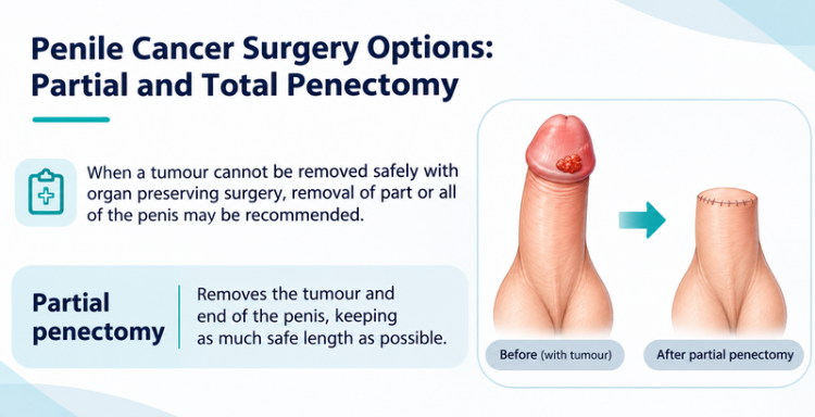
When a tumour cannot be removed safely with organ preserving surgery, removal of part or all of the penis may be recommended.

  • Partial penectomy: Removes the tumour and end of the penis, keeping as much safe length as possible.
  • Total penectomy: Considered when too little would remain for useful function. A new urine opening is made behind the scrotum (perineal urethrostomy), so urine is passed sitting down.

Many men still pass urine standing after partial penectomy, depending on remaining length. Sexual function depends on remaining length, sensation, erectile tissue, general health and emotional wellbeing. Counselling and psychosexual support can help with body image and relationships.

Penile Cancer Lymph Node Treatment: Assessment and Surgery

Penile cancer usually spreads first to the groin lymph nodes, whose status strongly predicts outcome, so node decisions are made separately.

Normal sized nodes may hold microscopic cancer, while enlarged nodes may reflect infection. Assessment is guided by examination and tumour risk:

  • Nodes not felt, higher risk tumour (T1b or above): Dynamic sentinel lymph node biopsy at an experienced centre, or a limited groin dissection.
  • Nodes not felt, very low risk tumour: Regular examination and ultrasound may be enough.
  • Nodes felt or abnormal on ultrasound: Needle sampling to confirm cancer. If sampling confirms cancer, treatment depends on the size, mobility and extent of the affected nodes. Operable disease may be treated surgically, while bulky or fixed nodes may require chemotherapy first.
  • Several positive groin nodes or cancer outside a node: Pelvic lymph node treatment may be added.

Groin surgery can cause leg swelling (lymphoedema), so its extent is matched to risk.

Advanced Penile Cancer Treatment: Chemotherapy and Radiation

By type of advanced disease:

  • Locally advanced tumour: Usually surgery; radiation or chemoradiation if surgery is unsuitable.
  • Bulky or fixed groin nodes: Cisplatin based chemotherapy before surgery can shrink disease and allow surgery in responders.
  • Extensive nodal disease found at surgery: Chemotherapy or radiation afterwards may lower the risk of return.
  • Distant spread: Platinum based chemotherapy first; clinical trials, or immunotherapy or targeted therapy for selected tumour markers, may be considered.

When disease is limited to the penis and regional nodes and can be completely removed, treatment may aim for cure, although the likelihood falls as nodal disease increases. With distant spread, the aims are usually disease control, symptom relief and quality of life.

Penis Cancer Treatment Planning: Tests and Individual Factors

Tests depend on the tumour and likelihood of spread:

  • Examination of the penis and both groins.
  • Biopsy and pathology review.
  • MRI or ultrasound when needed to clarify local tumour depth.
  • CT or PET CT when nodes are involved or the risk of spread is higher.

Not every patient needs every scan.

Alongside stage, the following factors influence treatment selection:

  • Tumour size, location, grade and other risk features.
  • Whether the penis can be safely preserved, and previous treatment.
  • General health and fitness for treatment.
  • Personal priorities, including urinary and sexual function.
  • Ability to attend close follow up.

Multidisciplinary review and shared decisions personalise the plan.

What Our Patients Are Saying

" Dr. Ramani was very professional in his approach. He spent a lot of time to discuss the problem and compassionately handled a very complicated case. He is always accessible and ready to go the extra mile to put the patient at ease. Highly recommended! "
Manish Maheswari
On Google

Follow-Up After Penis Cancer Treatment: Recurrence and Recovery

Follow up is most frequent in the first two years, when most recurrences appear, with examination of the treated area and groins and imaging when indicated. Schedules vary with the treatment received and recurrence risk.

Report any new lump, sore or bleeding at the treated area, groin or leg swelling, or difficulty passing urine.

Visits also review urinary, sexual and emotional wellbeing.

 
Let’s Discuss! Request a Call Now!
White Call Now!
 

About Dr. Anup Ramani: Penis Cancer Surgeon in Mumbai

Dr. Anup Ramani (MCh, DNB, MS, DNB) is a uro oncological surgeon in Mumbai with more than two decades of experience.

His practice treats prostate, kidney, bladder, adrenal, testicular and penile cancers, along with enlarged prostate and kidney stones. His penis cancer surgery service includes partial and total penectomy. Patients receive a written cost estimate, support with hospital admission and round the clock access to the care team, and international patients are also supported. When chemotherapy or radiation is needed, care is coordinated with medical and radiation oncologists.

Conclusion: Choosing the Right Penis Cancer Treatment

Stage is the starting point for penis cancer treatment, not the whole answer. Your biopsy, staging results, health and priorities together shape the most suitable plan, so review them with a qualified specialist.

Penis Cancer Stages and Treatment FAQs

Stage shows how far the cancer extends, guiding how much surgery and node or other treatment is needed.

Sometimes. Topical creams may treat selected non invasive lesions such as PeIN. Small invasive tumours may be treated with local excision, glans surgery or radiation in suitable patients. Laser treatment requires careful specialist selection.

Stage describes how far the cancer has spread. Grade describes how abnormal the cells look.

No. Swelling may be due to infection, and normal sized nodes can still hold cancer. Sampling or biopsy is needed to know.

When organ preserving surgery is not safe. Partial penectomy is used when enough healthy shaft can remain; total penectomy when it cannot.

For bulky or fixed groin nodes, chemotherapy can shrink disease so it can be removed completely.

In some cases, when cancer is limited to the penis and regional nodes and can be completely removed. With distant spread, treatment focuses on control.

Regular examination of the treated area and groins, most often in the first two years.

PARTIAL PENECTOMY
  • Partial penectomy is done in cases where glans and distal penis is involved with carcinoma. 
  • Partial penectomy is a type of organ-preserving surgery. Preservation of sexual and micturational function depends on the surgical dissection and reconstruction of residual urethra.
Kidney Stone Removal
  • Patients who develop stones in the kidney or ureter, often experience severe pain.
  • This condition usually needs a procedure to remove the kidney stones.
  • This procedure is called ureteroscopy and is performed very commonly.
  • It does not require any cuts and hence it is painless.
  • The procedure is performed with an endoscope inserted through the penis under spinal anesthesia.
  • The scope is inserted through the penis into the kidney and stones are dissolved with a laser.
  • The procedure takes about 40-50 minutes. 
  • A catheter (urine pipe) is kept after the procedure to drain the bladder. A stent is kept in the kidney at the same time.
  • Patient is mobile and walking in the room the same evening.
  • Hospital stay is one night and patient is discharged the next day after removal of the catheter.
  • Patient has to come back after six weeks to remove the stent in the kidney.
  • Patients can resume office a week after surgery and heavy activities like running, weight lifting, a month after the procedure.
  • We offer fixed packages for this procedure which can be obtained by calling our helpline +91 9967666060.
  • Men with an enlarged prostate, which is a normal ageing changes, often experiencing difficulty passing urine. This condition usually needs a procedure to trim the prostate and relieve the blockage.
  • This procedure is called TURP and is performed very commonly.
  • It does not require any cuts and hence it is painless.
  • The procedure is performed with an endoscope inserted through the penis under spinal anaesthesia.
  • The overgrown prostate is dissolved with a laser bloodlessly.
  • The procedure takes about 40 minutes.
  • A catheter (urine pipe) is kept after the procedure to drain the bladder.
  • Patient is mobile and walking in the room the same evening.
  • Hospital stay is two nights and patient is discharged with the catheter, which is removed after 4 days.
  • Patients can resume office a week after surgery and heavy activities like running, weight lifting, a month after the procedure.
  • We offer fixed packages for this procedure which can be obtained by calling our helpline +91 9967666060.
ROBOTIC ADRENALECTOMY FOR ADRENAL GLAND TUMOUR
  • Robotic adrenalectomy is a sophisticated, complex surgery and it is very important that an experienced surgeon performs this surgery to avoid major complications.

  • Once the anesthesia is done, and patient positioned, three micro cuts (3mm each) are made in the patient’s abdomen.

  • The arms of the Da Vinci robot are connected to the cuts via ports (tubes).

  • Dr. Ramani then sits in the controlling console to perform the surgery.

  • On an average, a robotic adrenalectomy takes one hour.

  • The surgery is almost completely bloodless and there has never been any need to transfuse blood after surgery.

  • A urine catheter and bag to drain the bladder is inserted during surgery.

  • A tiny drain pipe may be inserted in the surgical side of the abdomen, connected to a bag.

  • Patient is kept nil-by-mouth the day of the surgery, with IV fluids. Sips of water are started the next day and solid food by day three.

  • The drain pipe, if kept, is removed in the room on day 2 after surgery.

  • The catheter is removed on day two after surgery.

  • Total hospital stay for robotic adrenalectomy is 4 nights (including night before surgery).

  • Post discharge, a doctor from the surgical team visits the patient at home/ hotel room once every day.

RETURN TO ACTIVITY
  • On the day of discharge, patient is totally self-sufficient. They are able to walk freely without any pain, dress themselves, shower, toilet and they do not need to hire any nurse or help at home. Almost all patients are back to work within 2 weeks of surgery.

  • Heavy activities like running, weight lifting can be resumed after a month

FOLLOW UP AFTER SURGERY
  • Follow up after an adrenalectomy is in the form of CT scans, once a year for 5 years.
    Local patients usually meet Dr. Ramani after two weeks to discuss report.

  • Outstation patients are counselled on a phone consultation.

ROBOTIC SURGERY FOR BLADDER CANCER
  • Dr. Ramani is one of the very few surgeons in India who has the expertise to perform a robotic surgery for bladder cancer, which includes removing the urinary bladder and reconstructing a new bladder robotically.
  • Robotic radical cystectomy is an extremely sophisticated, complex surgery and it is very important that an experienced surgeon performs this surgery to avoid major complications.
  • Once the anaesthesia is done, and patient positioned, six micro cuts (3mm each) are made in the patient’s abdomen.
  • The arms of the Da Vinci robot are connected to the cuts via ports (tubes).
  • Dr. Ramani then sits in the controlling console to perform the surgery.
  • On an average, a robotic radical cystectomy with an ileal conduit takes 3-4 hours.
  • The surgery is almost completely bloodless and there has never been any need to transfuse blood after surgery.
  • A urine catheter and bag to drain the new bladder is inserted during surgery.
  • Two tiny drain pipe in inserted in the surgical side of the abdomen, connected to a bag.
  • Patient is kept nil-by-mouth for 4 days after surgery with IV supplementation of patient’s daily requirements of calories, fats, carbohydrates, proteins and electrolytes.
  • The drain pipes are removed in the room on day 3-5 after surgery.
  • Total hospital stay for radical cystectomy is 8 nights (including night before surgery).
  • Post discharge, a doctor from the surgical team visits the patient at home/ hotel room once every day.
RETURN TO ACTIVITY
  • On the day of discharge, patient is totally self-sufficient. They are able to walk freely without any pain, dress themselves, shower, toilet and they do not need to hire any nurse or help at home.
  • Almost all patients are back to work within 6 weeks of surgery. Heavy activities like running, weight lifting can be resumed after two months.
FOLLOW UP AFTER SURGERY
  • Follow up after a radical a cystectomy is in the form of CT scans, once a year for 5 years.

  • Histopathology report: Local patients usually meet Dr. Ramani after two weeks to discuss report.

  • Outstation patients are counselled on a phone consult. Depending on the report, patient may or may not need chemotherapy after surgery.

  • If chemo is needed, patients may choose to get it done with a medical oncologist of their choice or avail the services of one of the four medical oncologists on our team.

ROBOTIC RADICAL/PARTIAL NEPHRECTOMY FOR KIDNEY CANCER
  • Robotic partial nephrectomy is a sophisticated, complex surgery and it is very important that an experienced surgeon performs this surgery to avoid major complications. Robotic radical (total) nephrectomy is
  • relatively easier but still requires significant experience to consistently deliver results.
  • Once the anaesthesia is done, and patient positioned, five micro cuts (3mm each) are made in the patient’s abdomen.
  • The arms of the Da Vinci robot are connected to the cuts via ports (tubes).
  • Dr. Ramani then sits in the controlling console to perform the surgery.
  • On an average, a robotic radical nephrectomy takes one hour and a robotic partial nephrectomy takes about an hour and half.
  • The surgery is almost completely bloodless and there has never been any need to transfuse blood after surgery.
  • A urine catheter and bag to drain the bladder is inserted during surgery.
  • A tiny drain pipe in inserted in the surgical side of the abdomen, connected to a bag.
  • Patient is kept nil-by-mouth the day of the surgery, with IV fluids. Sips of water are started the next day and solid food by day three.
  • The drain pipe is removed in the room on day 3 after surgery. The catheter is removed on day two after surgery.
  • Total hospital stay for radical/partial nephrectomy is 4 nights (including night before surgery).
  • Post discharge, a doctor from the surgical team visits the patient at home/ hotel room once every day.
RETURN TO ACTIVITY
  • On the day of discharge, patient is totally self- sufficient. 
  • They are able to walk freely without any pain, dress themselves, shower, toilet and they do not need to hire any nurse or help at home. 
  • Almost all patients are back to work within 2-3 weeks of surgery.
  • Heavy activities like running, weight lifting can be resumed after a month.
FOLLOW UP AFTER SURGERY
  • Follow up after a radical/partial Nephrectomy is in the form of CT scans, once a year for 5 years.
  • Local patients usually meet Dr. Ramani after two weeks to discuss report. 
  • Outstation patients are counselled on a phone consultation.